
Panels
Four ready panels, one chip each: the question each one answers, what is on the chip, and how to read the result.
Four panels
A panel is a set of pre-conjugated ligands on fixed oligo addresses, usually with a built-in reference. The Immune Profiling panel uses all eight addresses for binders. Layout, replicates and controls are designed in advance, so a run can focus on samples and interpretation.
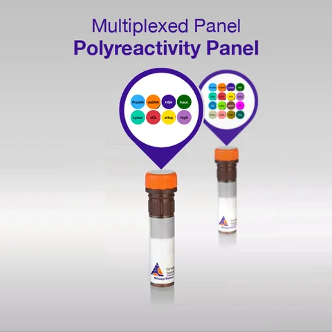
Polyreactivity Panel
Does the candidate bind surfaces it should not?
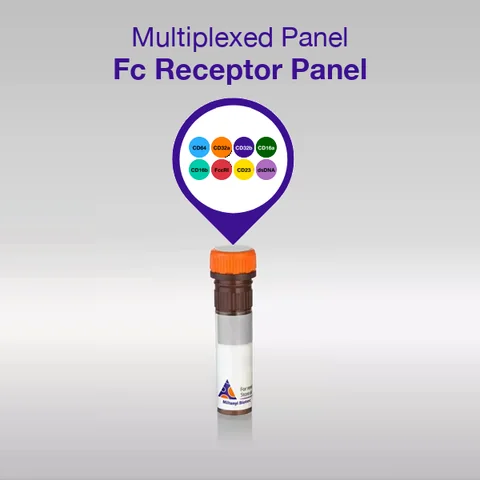
Fc Receptor Panel
How does the antibody engage immune effector cells?
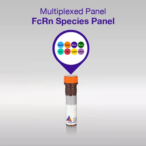
FcRn Species Panel
How long will it circulate, across species?
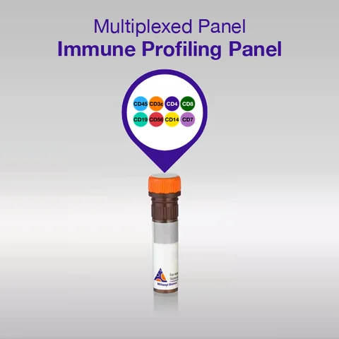
Immune Profiling Nanobody Panel
Which immune cell markers are present?
One chip, five steps
- 8-plex chip
- Oligo panel, pooled
- Loading
- Titration (SCK)
- Regeneration
Choose a panel, then play the five steps or step through them. The chip shows the real address map, and position n of the panel table sits on address n. Then switch the candidate and run the titration again.
In the animation, each address follows a 1:1 model. Protein A/G gets a high affinity, the other ligands a low one, so their response grows mostly at the top concentrations. The titration follows the polyreactivity runs on the developability page. The Fc Receptor, FcRn Species and Immune Profiling panels are not in the selector, because this page does not list which address carries which ligand.
Where the panels fit in antibody development
Developability questions usually need separate platforms. Here each is answered on one chip, with the intact antibody in a relevant matrix. Polyreactivity, Fc receptor and FcRn binding each have a panel; plasma protein binding is an assay that captures the antibody through anti-Fc.
Does it bind surfaces it should not?
Does plasma protein bind to it, and does it stay bound?
How does it engage immune cells?
How long does it circulate?
Polyreactivity Panel
Does the candidate bind surfaces it should not?
Why it matters. Polyreactivity is linked to fast clearance, a short half-life and attrition in clinical development (Jain et al., PNAS 2017). Endpoint assays report it without kinetics and on a separate platform; the panel measures it label-free, on the same instrument as the target kinetics.
Readout. One response per ligand, summarized as a Polyreactivity Index. The fingerprint across the ligands shows which chemistries a candidate binds, and whether the binding is broad or focused. Compare candidates within one run, under identical conditions.
Two formats. Essentials (8-plex, 8 replicates per ligand) for routine triage in every discovery cycle; Plus (16-plex, 4 replicates) for in-depth assessment ahead of lead nomination.
The ligands and why they are there
Each ligand stands for one kind of unwanted interaction: charge, lipid, glycan, plasma protein or nucleic acid. Positions 1 to 8 carry the same ligands on the same oligo addresses in both formats. An Essentials screen and a Plus follow-up therefore give directly comparable results.
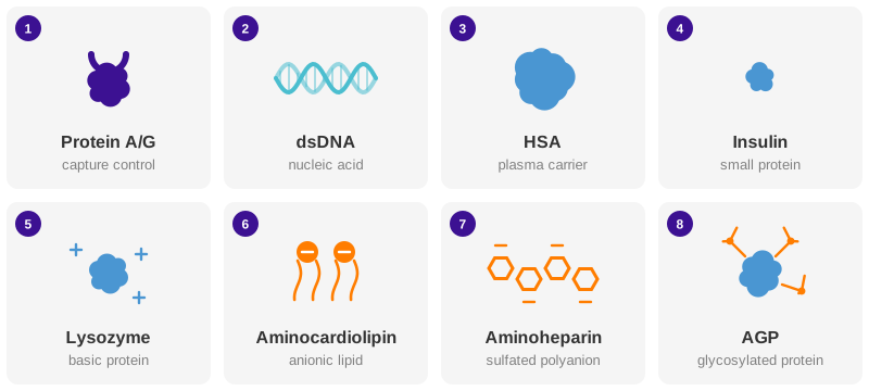
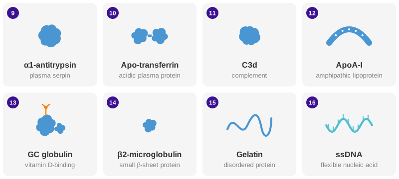
| # | Ligand | What binding to it reveals |
|---|---|---|
| Positions 1 to 8 · Essentials and Plus | ||
| 1 | Protein A/G | Control: confirms the antibody was captured and normalizes each injection. Not a liability axis. |
| 2 | dsDNA | Reference surface; also reports nucleic-acid binding, a classic polyreactivity signal. |
| 3 | Human serum albumin | Binding to the main plasma carrier protein. |
| 4 | Insulin | Binding to a small protein; a standard polyreactivity antigen. |
| 5 | Human lysozyme | Electrostatic binding to a basic, positively charged protein. |
| 6 | Aminocardiolipin | Binding to an anionic lipid, as found in cell membranes. |
| 7 | Aminoheparin | Binding to a sulfated polyanion; flags positively charged patches. |
| 8 | α1-acid glycoprotein | Binding to a heavily glycosylated plasma protein. |
| Positions 9 to 16 · Plus only | ||
| 9 | α1-antitrypsin | Binding to an abundant plasma serpin. |
| 10 | Apo-transferrin | Binding to an acidic plasma protein. |
| 11 | Complement C3d | Interaction with the complement system. |
| 12 | Apolipoprotein A-I | Binding to an amphipathic lipoprotein component. |
| 13 | GC globulin | Binding to a vitamin D-binding glycoprotein. |
| 14 | β2-microglobulin | Binding to a small β-sheet protein. |
| 15 | Gelatin | Binding to a disordered protein. |
| 16 | ssDNA | A second, flexible nucleic-acid surface. |
Fc Receptor Panel
How does the antibody engage immune effector cells?
Why it matters. The Fab binds the target; the Fc region engages Fcγ and Fcε receptors on immune cells and so decides on phagocytosis, ADCC and degranulation.
Readout. Kinetics and affinity for each receptor, read as a profile. The balance between activating and inhibitory receptors is more informative than any single value, because Fc receptor binding is weak, avidity-driven and glycan-dependent. Set the profile against cell-based assays such as ADCC and ADCP.
| Receptor | Signal | Drives |
|---|---|---|
| FcγRI (CD64) | activating | phagocytosis |
| FcγRIIa (CD32a) | activating | platelets, monocytes |
| FcγRIIb (CD32b) | inhibitory | damps the response |
| FcγRIIIa (CD16a) | activating | NK cells, ADCC |
| FcγRIIIb (CD16b) | activating | neutrophils |
| FcεRI | activating | mast cells, allergy |
| FcεRII (CD23) | regulatory | IgE feedback |
| Oligo only | reference | checks binding to the DNA layer |
FcRn Species Panel
How long will the antibody circulate, and how does that translate between species?
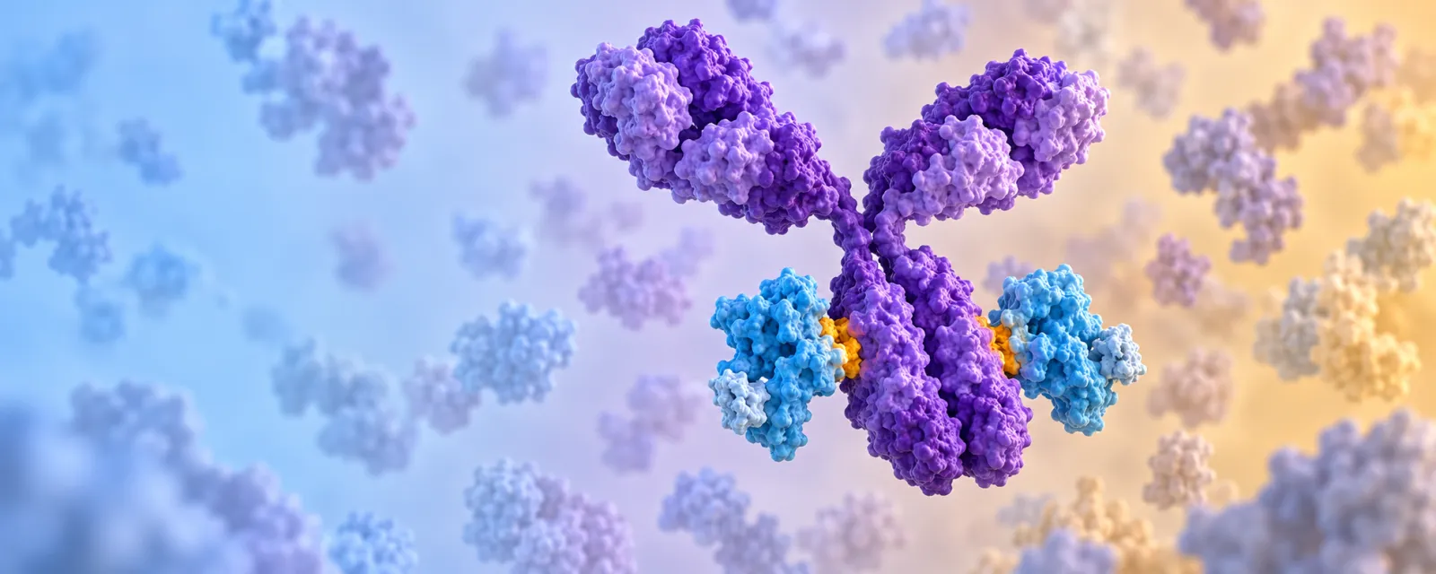
Why it matters. FcRn rescues IgG from lysosomal degradation. It binds in the acidic endosome at pH 6.0 and releases at the cell surface at pH 7.4. This switch sets the serum half-life.
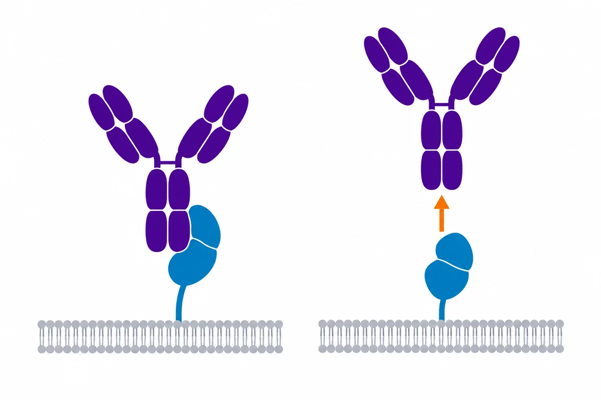
Readout. Kinetics at pH 6.0 and at pH 7.4, one single-cycle run each. Strong binding at pH 6.0 supports a long half-life; persistent binding at pH 7.4 is a liability, because the antibody is not released.
| On the chip | Role |
|---|---|
| Human FcRn | Clinical reference |
| Cynomolgus, marmoset FcRn | Primate models |
| Mouse, rat FcRn | Rodent models |
| Minipig FcRn | Large-animal model |
| Human FcRn L320A | IgG-binding-deficient specificity control |
| Oligo only | Reference for the DNA layer |
Immune Profiling Nanobody Panel
Which immune cell markers are present in a sample?
Why it matters. Eight markers resolve the main leukocyte classes: T cells with their helper and cytotoxic subsets, B cells, NK cells and monocytes.
Readout. Binding to each marker-specific nanobody, measured in parallel on one [Oligo|Oligo] 8-plex chip, in whole blood, PBMCs or serum-containing media.
| Marker | Identifies |
|---|---|
| CD45 | all leukocytes |
| CD3ε | all T cells |
| CD4 | helper T cells |
| CD8 | cytotoxic T cells |
| CD19 | B cells |
| CD56 | NK cells |
| CD14 | monocytes |
| CD7 | T and NK lineage |
At about 15 kDa the nanobodies pack densely on the ridges. With one binding domain and no Fc, they add no Fc-mediated background. Each binder comes in its own vial, on the oligo sequence you choose.