
DEL hit validation
Measure DNA-encoded library (DEL) hits on their DNA. Each hit finds its own molograms by its DNA, and you get kinetics for many hits in one run, before any off-DNA synthesis.
The question
Which of my DEL hits bind the target, how strongly and how fast, and do they bind anything else?
A DEL selection gives a list of enriched barcodes, not affinities. The next step is to decide which hits deserve the work of off-DNA synthesis. That decision is easier with measured kon, koff and KD for many hits, side by side. Two published papers show how this works.
The setup
| Chip | DNA chip. Each oligo address sits on its own molograms, so several compounds share one chip |
|---|---|
| Hit on the chip | The hit keeps its DNA. The Oligo Adapter 5′-Phosphate is ligated to the DNA of a hit; the Oligo Adapter Amine is for on-DNA resynthesis |
| Analyte | The protein flows over the chip. Study 1: carbonic anhydrase IX (CAIX). Study 2: fibroblast activation protein (FAP) and five off-target proteins. Study 3: VHL |
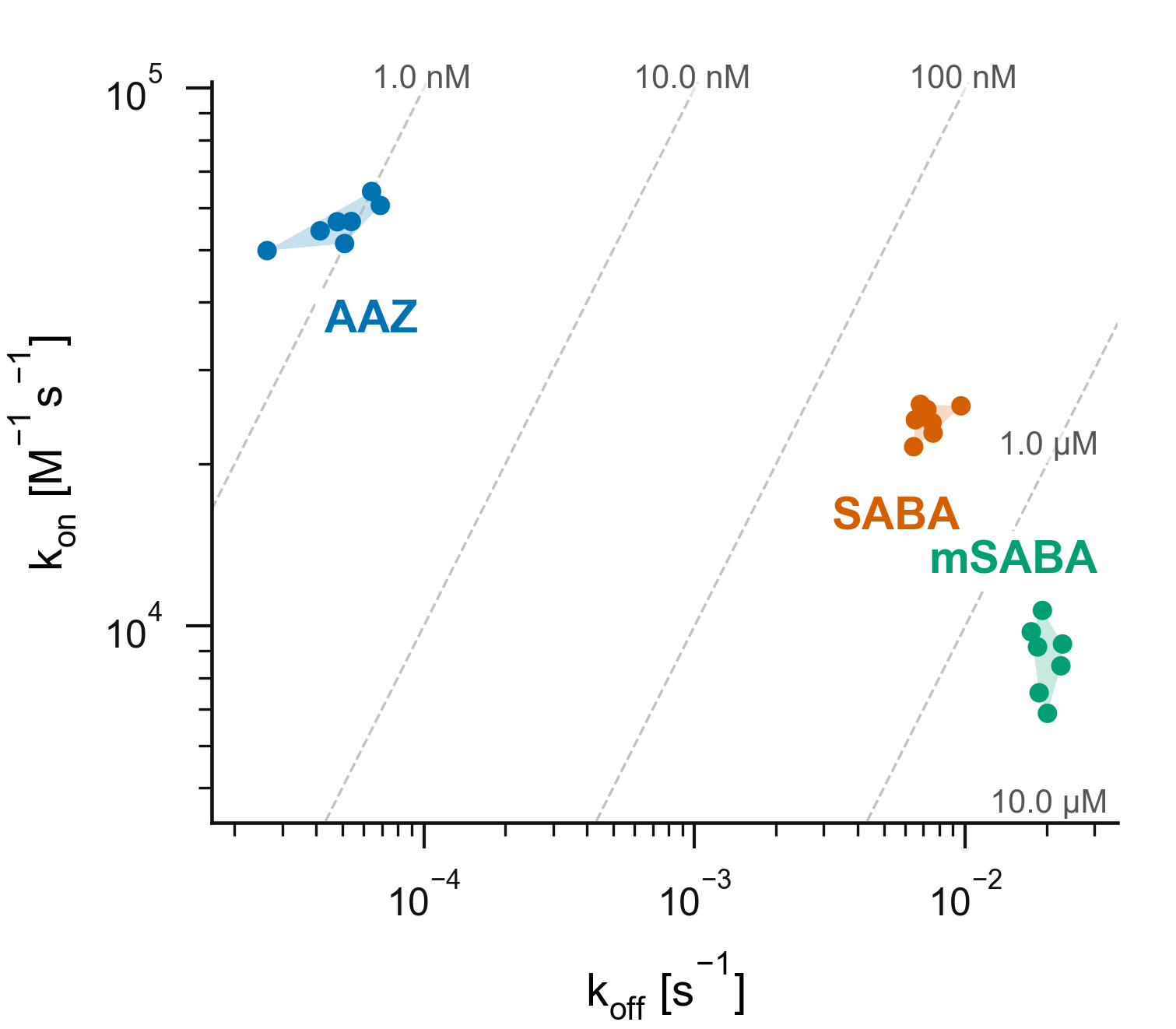
| Study 1: CAIX | The reference inhibitor AAZ and two DEL ligands, SABA and mSABA. CAIX in 11 steps from 0.36 nM to 7.1 µM, seven replicate molograms per compound |
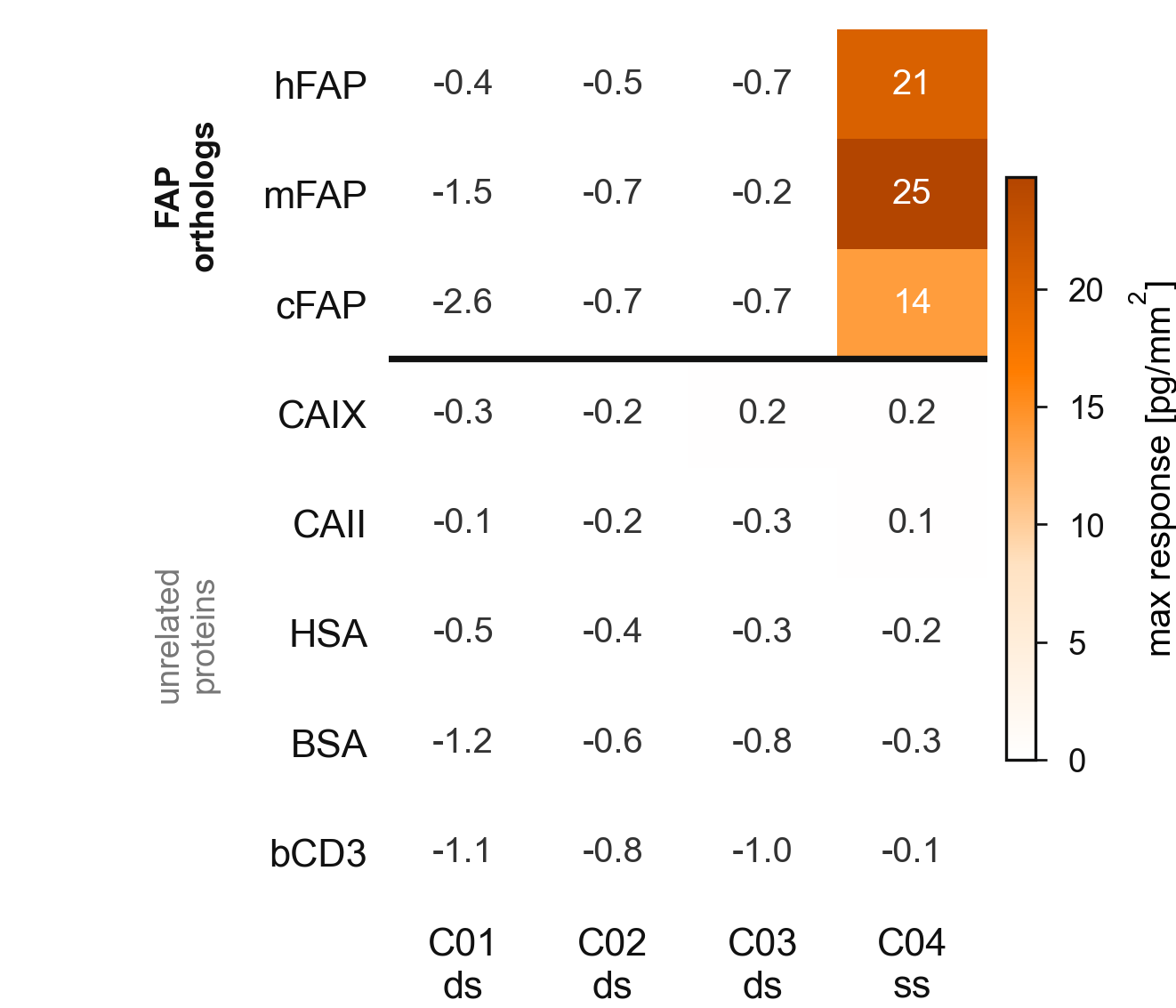
| Study 2: FAP | The FAP ligand oncoFAP on DNA, in four constructs on a DNA chip with a PEG background, 16 molograms each. Code C04: purified, with a single-stranded DNA end. Codes C01 to C03: crude ligation products with a double-stranded DNA end. FAP from human (hFAP), mouse (mFAP) and dog (cFAP), 195 pM to 200 nM. CAIX, CAII, HSA, BSA and CD3 as off-target proteins, up to 100 µM |
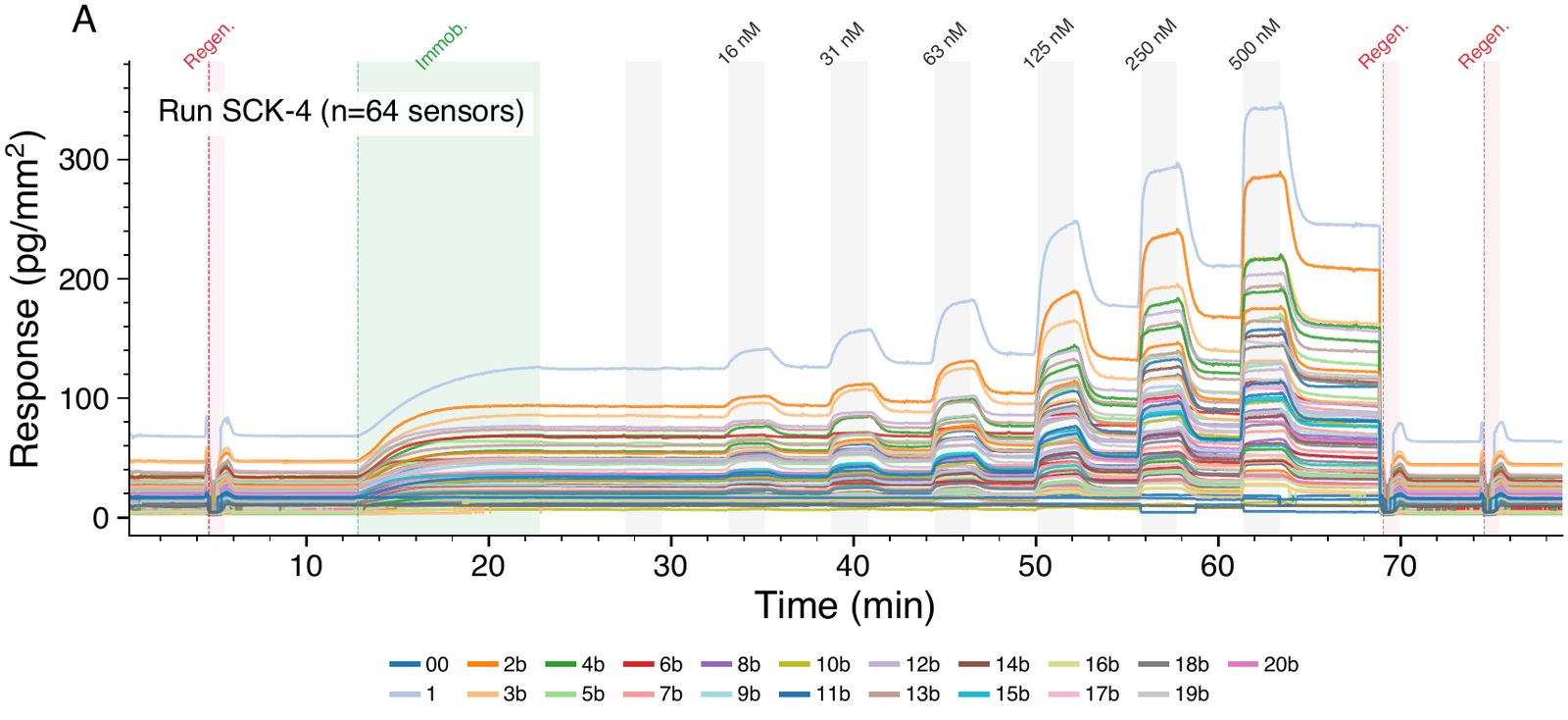
| Study 3: VHL | 20 DNA-VHL ligand conjugates (DNA-PROTACs) that differ in their linker, pooled and immobilized together. Each takes three molograms. VHL from 16 to 500 nM |
| Assay | Single-cycle kinetics (SCK): rising concentrations without regeneration in between |
Eight hits on an 8-plex DNA chip are shown, eight molograms each. Play the workflow or step through it.
Measured result
CAIX: two DEL ligands ranked against a reference inhibitor
FAP: on-target, cross-species and off-target binding
VHL: 20 DNA-PROTAC linkers in one run
How to read it
- The DEL ligands are clearly ranked. Replicate KD values on CAIX are 264 to 375 nM for SABA and 1.8 to 2.9 µM for mSABA. The reference AAZ gives 0.53 to 1.14 nM.
- Kinetics show why. AAZ dissociates about 100 times slower than SABA. SABA binds faster and dissociates slower than mSABA.
- Replicates agree. The seven fits of each compound form a tight group, well apart from the other two compounds.
- The FAP ligand binds all three orthologs. Human, mouse and dog FAP give 14 to 25 pg/mm² on the purified construct. The paper reports KD of 0.66 to 1.87 nM across the orthologs for the PEG4-C6 linked construct, 16 molograms each.
- No off-target binding. CAIX, CAII, HSA, BSA and CD3 give no response up to 100 µM, close to zero in every column.
- The DNA format matters. The constructs with a double-stranded DNA end do not bind FAP at all. Enzyme inhibition and fluorescence polarization assays in the paper agree.
- One run covers a whole series. All 20 conjugates bind VHL in the same run, each on its own molograms. The blank address shows no binding.
- The linker changes affinity. Mean KD on VHL ranges from 293 nM (compound 19b) to 1.15 µM (compound 6b), from 8 to 12 fits each. Compounds with a higher cLogP bound more tightly (Pearson r = −0.74 for cLogP against log KD, n = 20).
- What it does not show is how the same molecule binds without its DNA. These are on-DNA values. The VHL study measures binary binding only, not ternary complex formation or degradation in cells.
Why it works
Each mologram carries one oligo sequence on its ridges, so a hit with the matching DNA binds only to its own molograms. The protein is the analyte, so the signal comes from the protein mass that binds the hit, not from the small molecule itself. The readout counts only mass that binds in the line pattern of the mologram.
Read more: capture chemistry on ridges, matched grooves, what sets the detection limit, and from focal spot to coherent mass density.
More
- Application: DEL hit validation and targeted protein degradation
- Documentation: DEL ligation protocol
- Paper: Raschke, Notova, Gatterdam, Frutiger and Brunschweiger, RSC Chem. Biol. 2026, doi:10.1039/d6cb00011h
- Paper: Favalli et al., High-throughput binding kinetic measurements of DNA-encoded library-derived hits by focal molography, J. Med. Chem. 2026, doi:10.1021/acs.jmedchem.6c01097
- App note: Fast and convenient hit validation for DNA-encoded libraries (2024)
- App note: One on-DNA ligand against many targets: selectivity profiling for DEL hit validation (2026)
- Products: DEL oligo adapters and DNA chips
- Training: The software: from design to report