
Serum, plasma and lysate
Measure binding in the sample the molecule will meet: undiluted serum, plasma or cell lysate, without purification or dilution.
The question
Does my binder still bind in serum, and with the same kinetics as in buffer?
Binding measured in buffer can differ from binding in blood or lysate. Matrix proteins compete for the ligand, stick to it, or lower the free analyte concentration. SPR and BLI also support complex-sample assays. Their reference surfaces, matrix blanks and other corrections address background according to the instrument and assay. Dilution or purification can change the interaction context, so choose sample preparation to fit the biological question. See the method comparison and primary sources.
The setup
| Chip | DNA chip; the ligand reaches its molograms by DNA-directed immobilization (DDI) |
|---|---|
| Ligand | An anti-GFP nanobody (VHH) on an oligo. The same VHH is sold as a capture adapter for GFP-tagged proteins |
| Analyte | GFP, 125 pM to 32 nM |
| Assay | Single-cycle kinetics (SCK), after five blank injections of serum |
| Sample | Undiluted human serum. The same titration in PBST buffer, for comparison |
Measured result
How to read it
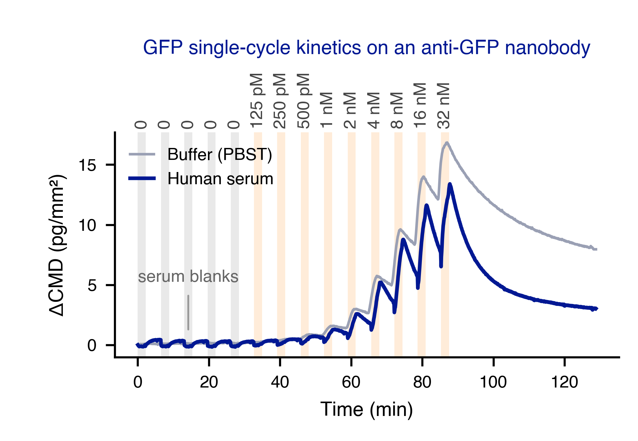
- The serum blanks stay flat. Undiluted serum alone adds less than 0.5 pg/mm² of coherent signal on these molograms.
- GFP binds in serum. Each concentration step raises the serum trace, as it does in buffer.
- Serum changes the observed response. At the highest concentrations the serum response is lower and dissociates faster than in buffer. Measuring in the sample shows this difference directly; a buffer-only experiment cannot.
- What it does not show is the cause of the difference. Competition, non-specific interaction with GFP and a changed free concentration all fit. A non-binding control ligand and matrix-matched blanks help separate them.
Why it works
The MACS Matchmaker reads coherent mass density: only mass that binds in the line pattern of the mologram builds the signal. Serum proteins that adsorb equally on ridges and grooves add no coherent signal, so no reference surface has to be subtracted. Matched grooves keep the adsorption equal in crude samples.
A cross-section through four ridges and four grooves of one mologram. Serum flows over it, first alone as a blank, then with GFP. The traces are a model. On the measured chip above, the serum blanks added less than 0.5 pg/mm² of coherent signal. Then set the grooves lighter or heavier than the ridges and play it again.
Read more: why shared changes cancel, how backfilling matches the grooves, and reading CMD together with the refractometric readout MD.
More
- App note: Context Matters (May 2024)
- Antibodies binding plasma and serum proteins: developability
- Training: Measure in serum, plasma, and lysate
- Documentation: Backfilling and NSB suppression